The first (thumb) finger's valgus deformation is an orthopedic pathology in which the finger is deformed, different.Mostly, the disease occurs in women after 30 years.Valgus deformation is perceived in 3% of people aged 15-30, 9% in those who are over 31-60 and 16% over 60.Not only does the problem cause strong aesthetic discomfort, it also causes changes in all leagues, in tendons, joints and legs.

Pathogenesis
With the valgus deformation of the first finger, the angle between the first and the second bone will be plus, and the first metatarsal bone will be observed.The thumb of the foot moves outward and the bone head pulls out, forming a bone.This bone does not allow the thumb to meet the norm and gradually differ outward.Tubercle (bone) prevents it from wearing shoes, causing inflammation, friction, and leads to inflammation of the bag of the first metatarsofalangeal joint.Gradually the bone swells and becomes painful.The inadequate position of the common causes early wear, cartilage lesion and the increase in bone growth.This, in turn, provokes the trauma of the foot and the further development of the pathology.As the disease progresses, the thumb moves the remaining fingers of the foot, leading to deformation in the form of a hammer.With the onset of the disease, the arthrosis of the plusnoplange joints, inflammation of the arthritis, finger joints, chronic inflammation of the first metatarsophalangeal joint, first metatarsal bone, flat pedas (chronic and transverse), bones and cartilage.
Shapes
In normal condition, metatarsal bones are parallel to each other.For some reasons, the first metatarsal bone is different, and as a result, a small bump appears on the leg, ligaments and tendons lose their elasticity and form their dysfunction.
The disease passes through several stages:
- Early.At this stage, the leg thumb has a difference of less than 15 degrees.
- Average.The first finger is deviated from 15 to 20.At the same time, deformation of the second finger can be observed.It rises above the thumb and becomes like a hammer.
- Difficult.The thumb has a difference of 30 degrees.All the fingers of the foot have already been deformed and the first Phalanx bottom has a high bone increase.In places where the foot is high, rough corn appears.
Reasons
The main provocative factor for the appearance of valgus deformation is irregular shoes.If a person wears tight shoes with a narrow toe or high rodent, the fingers are constantly in a poor (compact) position, which contributes to the development of valgus deformation.But not only this will be the cause of the disease.The following reasons affect the appearance of pathology:
- Traumatic damage to the lower leg and foot;
- Angle;
- Brain paralysis;
- Polyneuropathy;
- the low arc of the foot;
- flat foot;
- Congenital weakness of the muscle giggle;
- Chronic arthritis triggered by psoriasis;
- arthritis;
- Multiple sclerosis (accompanied by damage to the nerve fibers);
- Diabetes mellitus;
- gout (deposition of your lord in the tissues of the body);
- Hyper of joint mobility observed during Marthans and Down syndrome;
- youthful leg (rapid growth of foot size during teenage periods);
- Chat-Mari-Tuta disease (hereditary neuropathy, atrophy of the muscles of distal limbs);
- osteoporosis;
- Stop Overstrain on professional activities (waiters, athletes, ballerinas).
In the presence of provocative factors, the disease is moving rapidly.
Symptoms

Symptoms of pathology depend on the degree of foot damage.In the first phase, the redness of the redness, the redness of the bones, the pain in the fingers of the fingers increases during the walk.In the average stages of the disease, pain, swelling, bone growth, plus bone, dry corn of the finger is the middle phalanx under dry corn.Severe, exhaustive pains appear in the heavy stage of the foot and thumb.Dry corn is formed and the keratinization of the skin appears on the second and third walls of the fingers.
Diagnosis
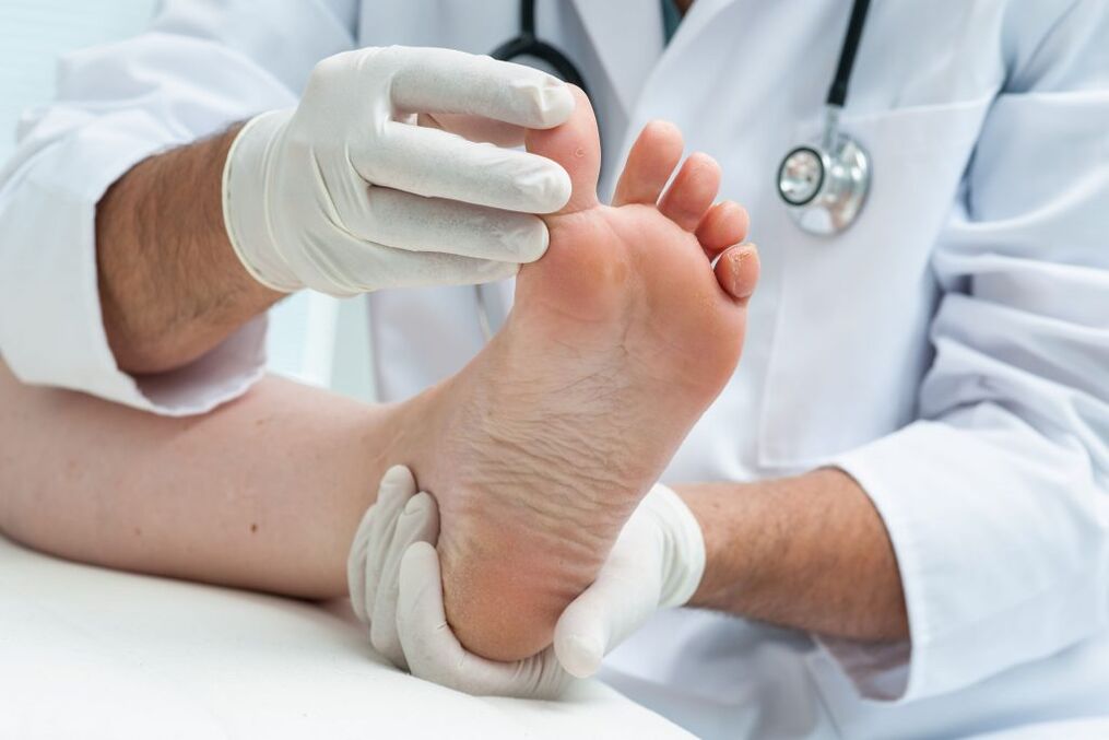
Diagnostic measures begin by collecting the patient's anamnesses.The doctor asks him to be disturbing symptoms, he cares about what provokes their appearance (foot load, walking, crowded shoes), there are metabolic, systemic diseases in the medical history, lower limbs injuries, hereditary diseases of the bones.An external inspection is then invited to ask a person.The doctor monitors the patient's walk while determining the intensity of the pain.The position of the leg thumb is examined, its location relative to other fingers, exploring the range of bending and extending it.Other external symptoms should be checked (swelling, redness, swelling under the fingers of the fingers, thickening of redness).The leg foot is carried out in three projections to determine the dislocation of the leg deformation, joint and related pathologies.The ultrasound of the vessels is performed to exclude the circulatory disorders of the legs.
Treatment
The therapy for the valgus deformation of the leg thumb is conventional and functional.In the early stages, when you can do it without surgery, doctors advise you to choose comfortable and correct shoes.You should not like your legs, friction.Comfortable shoes slow down the progress of the disease.However, the doctor advises you to buy special orthopedic devices:
- seals for the thumb joint bag (eliminates shoe pressure);
- Intodassen rollers, spacers that contribute to the proper distribution of the load on the leg.
All orthopedic devices reduce pain, but it is impossible to completely get rid of discomfort with their help.Corticosteroid injections are used to eliminate pain and inflammation, non -steroid anti -inflammatory drugs.To relieve cramps and restore joint mobility, massage and special gymnastics require to relax the legs.
The effectiveness of physiotherapy
Physiotherapy plays an important role in treating valgus deformation.To eliminate the disease, doctors write electrophoresis with calcium, hydrocortisone phonophoresis, paraffin and ozokeritic applications.One of the most effectiveMethods to eliminate the deformation of the foot thumb are the shock wave therapy (UVT).This procedure has a short -term effect on the painful zone with acoustic low -frequency pulses.With UVT, the symptoms of pain are eliminated and the effects on the factors contributing to their appearance are eliminated.The procedure is carried out by a special shock -wave device.Only pathological areas are affected without affecting healthy tissues.Ultrasound is used to improve metabolism processes.UVT is not performed in the presence of neurological, infectious, oncology, heart, somatic disease, diabetes mellitus, blood coagulation disorders.Shock wave therapy is not prescribed for pregnant women, breastfeeding women under the age of 18 and children.The effect of the procedure is observed after several procedures.The pain disappears, it can be easier.But it is impossible to completely remove the protruding bone using a UVT procedure.The doctor may prescribe at least 5-7 occupations.Depending on the stage of the disease, the procedure for performing procedures is also required.Some people are prescribed daily sessions, others will be sufficient for a weekly procedure.The result of UVT treatment lasts for a long time.
Surgery
The average and severe deformation of the foot thumb requires surgical treatment.To eliminate pathology, many methods have been developed to achieve the following goals:
- eliminate the bursitis of the first finger of the foot;
- Bowing the muscles around the injured joint to prevent a decline in the disease;
- Complete the reconstruction of the thumb bones.
If the deformation is not expressed heavily, the growth of joint bag growth through a small incision.After surgery, people need rehabilitation and must wear a bandage or shoe, using the foot switch, physiotherapy (at least 6 procedures).Currently, the following operations are performed with the Valgus deformation of the thumb:
- Small deformation correction.Small cuts are made on both sides of the thumb, through which miniature mills use the position of the metatarsal bones and the phalanx of the finger.
- Chevron-oosteotomy.The operation is performed with a difference of up to 17 degrees.Growth is cut out and the phalanx of the thumb is fixed with a screw and titanium wire.After a while, the structure is removed.
- Scarf oosteotomy.The operation is performed when the thumb is 18-40 degrees.During the intervention, manual exposure, the position of the metatarsal bones is corrected and then secured with two titanium screws.
The protruding bone of the leg thumb is not only ugly from aesthetic point of view, but also causes a lot of discomfort when walking.The disease, which has not yet completed treatment, leads to complications in the work of the entire muscle bone system.Therefore, everything should not be allowed on the gear and contact an orthopedic if the first signs of deformation appear


